Purpose
While growing liquid cultures in baffled flasks is an effective way to get large volumes of dense cell cultures, flasks can be unwieldy in high-throughput experimental workflows. Higher-throughput formats such as 24- and 96-well plates and test tubes can inhibit uniform mixing of cultures, often resulting in nutrient-deprived cells collecting at the bottom of wells.
While beads are primarily used for plating or lysing cells, we wondered if a single bead at the bottom of a well or test tube in a shaking incubator would create gentle turbulence similar to a stir bar, preventing sedimentation. The addition of beads to Schizosaccharomyces pombe (S. pombe) cultures vastly increased cell density across different vessels and culture volumes without increasing evaporation. Cultures with beads also produced cells that were larger and more homogeneous in morphology.
We conclude that adding beads to cultures improves culture quality and facilitates higher-throughput testing of S. pombe. We hope this finding is useful to others working with S. pombe, and would love to hear if anyone tests this approach for other organisms.
Background and goals
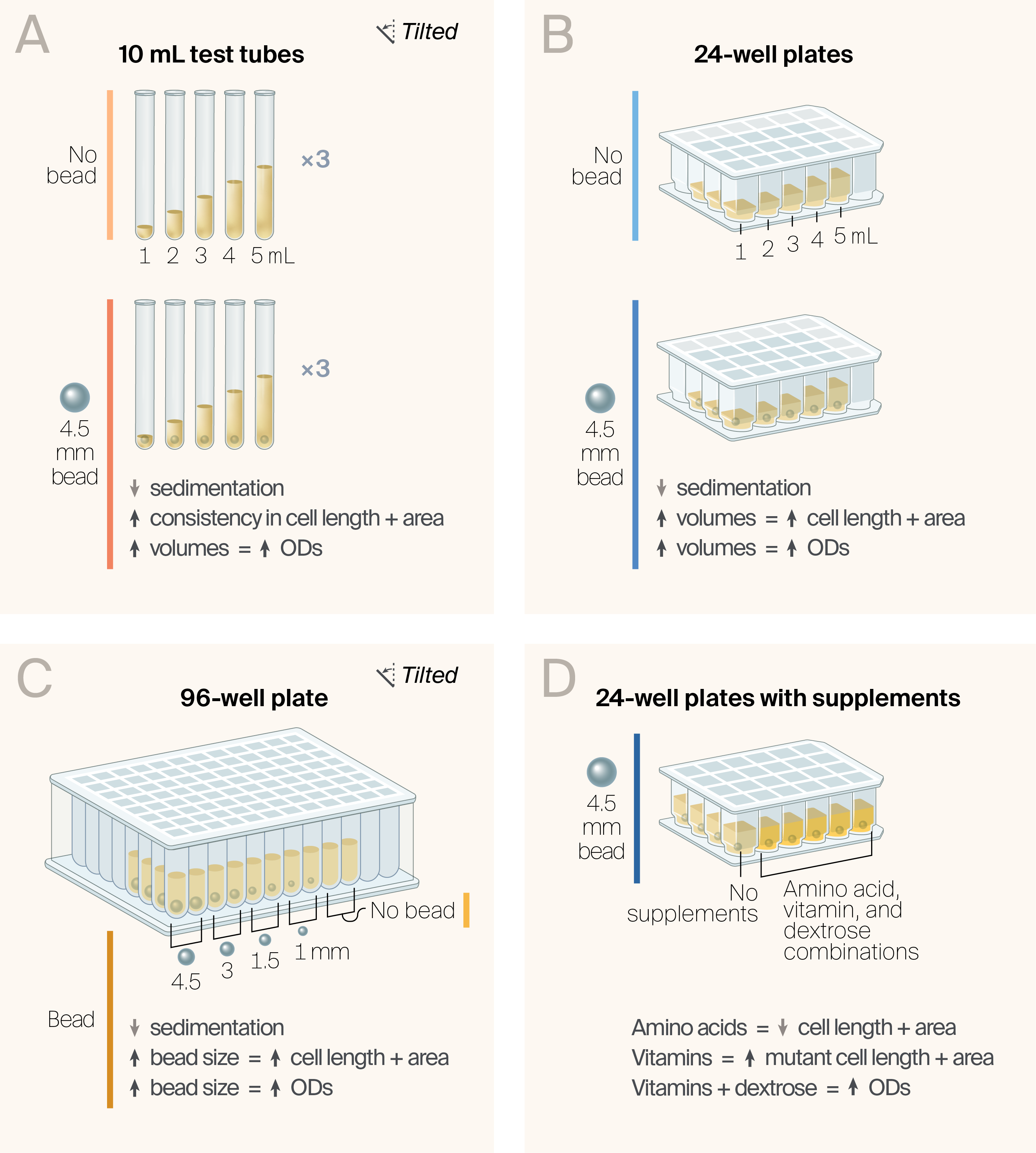
Figure 1. Beads prevent sedimentation across different growth conditions.
We set up experiments to test the effects of various growth variables on fission yeast growth. We found that 4.5 mm beads (A) in test tubes, (B) in 24-well plates, and (C) in 96-well plates prevent precipitation and yield more consistent cellular phenotypes, especially at higher volumes.
(D) Supplementation with different common EMM media supplements has different effects, with amino acids decreasing cell length and area, and both vitamins and dextrose increasing cell length and area.
We're testing S. pombe as a model organism for studying human disease. Central to this mission is finding reproducible cellular phenotypes with a high dynamic range between mutants and wild-type, allowing us to quantify how well candidate therapeutic molecules reverse mutant phenotypes.
One prominent phenotype that reappears with different S. pombe strains and growth conditions is larger, elongated cells 1. However, length and volume phenotypes are often lost or obscured when cells enter the stationary phase 2, which occurs when liquid cultures oversaturate, settle, or have reduced access to nutrients. Our ideal culture conditions would promote log growth and homogeneity of cell phenotypes within a single culture.
While growing cultures in baffled flasks allows cells to grow without settling, this method is unwieldy for high-throughput experiments. Flasks take up lots of space in shaking incubators and are incompatible with automation and multi-channel pipettes. Additionally, because we do most of our experiments in Edinburgh minimal medium (EMM), which contains only essential nutrients, strains often take longer to grow, compounding the problem. We therefore set out to find the best methods for growing cells in plates or test tubes without settling and associated stationary-phase phenotypes.
Stir bars inspired us to try adding beads to our cultures. While shaking incubators can generate sufficient turbulence in large containers to prevent cell sedimentation, laminar flow dominates in narrow or tall containers, preventing turbulent mixing of layers — even at high rotation speeds. While small beads are primarily used in liquid cultures to lyse cells, these usually require detergents and work by vigorously vortexing the cultures to cause the beads to knock against each other. In contrast, using a single, large bead in liquid culture wells in a shaking incubator should decouple the movement of the well from that of its contents, creating gentle turbulence that prevents cells from sedimenting.
In this pub, we set out to answer the following questions for S. pombe:
- Can beads prevent cell cultures from sedimenting?
- Can beads improve culture growth speeds, densities, or cellular phenotypes?
- Do beads lyse or stress cells?
- Which vessels are the best for growing cells?
- Which bead size and culture volumes are best for growing cell cultures?
The approach
Our general strategy was to systematically vary key components of our culture system (bead presence, bead size, vessel type, and nutrient content in the media) and quantify indicators of culture growth/cell health (cell density and cell morphology) (Figure 1). We then looked for significant differences in our data to see which variables are associated with faster, more homogeneous cell growth.
Yeast strains
The Schizosaccharomyces pombe strains we used in this study were diploid and derived from the SP286 (wild-type) genetic background (h+/h+ ade6-M210/ade6-M216 ura4-D18/ura4-D18 leu1-32/leu1-32). The gene-deletion strain dea2Δ (mutant) was generated in the SP286 background. We obtained the strains used in this study (Table 1) from Bioneer. We transformed both strains with the non-integrating plasmid pSLF172 (ATCC, 87609) for growth on EMM −Uracil selection media.
| Name | Strain ID | Genotype | Ploidy | Selection markers | Genetic background |
|---|---|---|---|---|---|
| SP286 (wild-type) | SP286 + pSLF172 | Wild type h+/h+ ade6-M210/ade6-M216 ura4-D18/ura4-D18 leu1-32/leu1-32 | Diploid | -Uracil | SP286 |
| dea2Δ (mutant) | SPBC1198.02 + pSLF172 | dea2Δ SP286 | Diploid | -Uracil | SP286 |
Table 1. Strains used in this study.
We used a wild-type and a mutant strain in this study because they have very different baseline phenotypes. However, the effect of growth with beads was consistent between strains. Therefore, we only show results for wild-type. Data for the mutant are available on Zenodo.
EMM with adenine and leucine
We prepared Edinburgh minimal medium (EMM) with 32.2 g/L EMM powder (MP Biomedicals, 114110012-CF), 225 mg/L adenine (Ade) (Thermo Fisher Scientific, 163631000), and 225 mg/L leucine (Leu) (Thermo Fisher Scientific, L8912), and autoclaved. For vitamin-supplemented media, we added 2 µg/L folic acid (Ambeed, A133193), 200 µg/L riboflavin (Ambeed, A454767), and 400 µg/L pyridoxine (Sigma-Aldrich, P5669), then filter-sterilized. We used dilute NaOH (Sigma-Aldrich, 567530) to dissolve the folic acid, and then filter-sterilized it. For the amino acid-supplemented media, we added 225 mg/L L-histidine (Sigma-Aldrich, H8000), 225 mg/L L-lysine (Sigma-Aldrich, L5501), and 225 mg/L L-arginine (Sigma-Aldrich, 1.01587), then filter-sterilized. For the dextrose-supplemented media, we added 10 g/L of filter-sterilized dextrose (Sigma-Aldrich, PHR1000). For EMM agar plates, we added 18 g/L agar powder (Sigma-Aldrich, A5306) and autoclaved the media. We then poured 25 mL of media into each plate. We diluted all supplemented and unsupplemented media to a final volume of 1 L with ultrapure water.
General growth
For all treatments, we streaked both S. pombe strains on EMM agar +Ade +Leu selection plates and incubated for 2–7 days at 30 °C 2. We then took about half of a colony from each plate and diluted it in 500 µL of liquid EMM +Ade +Leu. From here, we added 10 µL of this dilution per 1 mL of media into each well and test tube. We performed the 24-well and test-tube experiments three days after the 96-well plate experiment, so we used different colonies and aliquots from the same plate. We grew all colonies overnight at 30 °C and 200 rpm in an Infors HT Multitron Pro shaking incubator.
Tracking culture growth in different vessels
Test tubes
We grew cultures in 13 mm test tubes (Avantor, 47729-572) for one day, measuring optical density (OD) at 595 nm in the morning and afternoon, and performing microscopy in the morning. We performed three biological replicates per genotype, bead presence, and volume. We grew cultures in tubes tilted at 40° on tube holders (NIH 3D, 3DPX-023347) at 30 °C. Test tube caps allowed airflow. We filled tubes with 1, 2, 3, 4, or 5 mL of liquid media. Two sets of 15 had beads in all tubes, while the other two had none. We inoculated two sets of 15 (one with beads and one without) with SP286 + pSLF172, and inoculated the other two with dea2Δ + pSLF172. This resulted in 60 test tubes in total.
3D-printing files for our tilted test tube holder base and test tube holder are available on NIH 3D (3DPX-023347).
24-well plates
We grew cultures in 24-well plates at 30 °C (Agilent, 202061-300) for one day, measuring OD values in the morning and afternoon, and performing microscopy in the morning. We performed three biological replicates per genotype, bead presence, and volume. We didn’t grow plates on tilted bases, since the wide bottoms of 24-well plates allow for ample room for 4.5 mm beads (Zymo Research, 50-444-634) to create turbulence in the cultures. We covered the plates with a gas-permeable plate seal (Sigma-Aldrich, A9224). We filled wells with 1, 2, 3, 4, or 5 mL of liquid media. Two plates had beads in all wells, while the other two had no beads. We inoculated two plates (one with beads and one without) with SP286 + pSLF172, and inoculated the other two with dea2Δ + pSLF172. These growth conditions and replicates resulted in 60 experimental wells. We didn't control for edge effects on 24-well plates. We also grew media-supplemented experimental wells on 24-well plates using the same plate growth conditions.
96-well plates
We grew cultures in 2 mL 96-well plates (Thermo Fisher Scientific, Z717274) for 2 days instead of 1, measuring OD values after the first night of growth and taking microscopy measurements on day 2. We grew eight biological replicates per genotype and bead size. We grew plates on bases tilted at 40° (NIH 3D, 3DPX-023346). We covered plates with a gas-permeable plate seal. We filled all wells with 1 mL of media. Beads varied every two columns in the following order: 1 mm, 1.5 mm, and 3 mm Zirconium beads (Grainger, 39P088, 39P089, 39P090); then 4.5 mm glass beads; and finally, no beads. We inoculated the first four rows with SP286 + pSLF172, and the last four with dea2Δ + pSLF172. These growth conditions and replicates resulted in 80 experimental wells. We avoided wells in columns 1 and 12 to control for edge effects, which are most pronounced in 96-well plates.
3D-printing files for our tilted 96-well plate base are available on NIH 3D (3DPX-023346).
Tracking culture growth with different media supplements
We autoclaved all EMM media at 90% of the final volume, allowing us to add sterile liquid vitamins, amino acids, and dextrose supplements to the media without diluting the media, then add water to reach 100% volume. We made 40% dextrose, 100× amino acid, and 1,000× vitamin aliquots. We added a single 4.5 mm glass bead and 5 mL of media into all wells on two 24-well plates. We inoculated one plate with SP286 + pSLF172 and another with dea2Δ + pSLF172.
We prepared eight different experimental supplement treatments:
- Samples 2, 5, 7, and 8 contained dextrose up to 3% (w/v)
- Samples 3, 6, 7, and 8 contained amino acids
- Samples 4, 5, 6, and 8 contained a mix of vitamins
- Sample 1 served as a no-supplement control
We performed three biological replicates for each supplementation and genotype. This resulted in 48 wells in total.
OD measurement
We added 100 µL of undiluted liquid culture to 96-well clear-bottom plates with lids. We loaded these into a SpectraMax iD3 plate reader at 30 °C and performed a five-sample well scan to measure the cultures' absorbance at 595 nm. We shook the plate at high intensity in a double orbital before the first read to homogenize cultures.
To process the data, we converted absorbances into OD values in Microsoft Excel using a linear calibration:
We derived this formula by comparing plate reader measurements to OD measurements that we obtained using cuvettes and a NanoDrop OneC spectrophotometer (Thermo Fisher Scientific, ND-ONE-W). 0.0753 is the average baseline absorbance of EMM media, and 0.0766 represents the slope of the linear regression, which represents the relationship between OD values measured in a cuvette using a Nanodrop and ABS values measured in the plate reader for the same cultures. It was valid for OD values below ~7. We then averaged these five OD-converted technical replicates to determine the OD for each well. Finally, we averaged the OD values again across all three biological replicates to get the average OD per treatment.
Microscopy
We added 75 µL of culture from each well or test tube to a single well of a 96-well glass-bottom microscopy plate. We imaged cells using DIC and a 40× objective on a Yokogawa CSU-W1 SoRa spinning-disk confocal microscope on a Nikon Ti2-E inverted microscope, fitted with an ORCA-Fusion BT digital sCMOS camera (Hamamatsu) and a LIDA Light Engine (Lumencor) for illumination, controlled with NIS-Elements software (v5.42.03). We acquired images using a Plan Apo λ 40× air objective (NA 0.95) with the 1.5× relay zoom engaged. Additionally, we used auto-navigation and auto-focus for image acquisition. We captured nine images per well with a 1024 × 1024-pixel region of interest and an exposure time of 200 ms.
Sedimentation imaging
Using the iPhone SE's rear camera, we imaged the bottoms of test tubes and wells in 24- and 96-well plates. These allowed us to qualitatively assess sedimentation in cultures. Representative sedimentation images of wild-type yeast under different culture perturbations are available in this GitHub repo.
Statistical tests
We determined statistical significance using Holm–Bonferroni-corrected Welch's t-tests, where *p < 0.05, **p < 0.01, and ***p < 0.001. For the experiments in Figure 2, Figure 3, and Figure 4, beadless cultures served as the control condition. For the experiments in Figure 4, beadless cultures served as the control condition across all bead sizes. For the experiments in Figure 6, cultures without supplements served as the control condition.
Image processing and analysis
We processed images using a custom pipeline built on arcadia-microscopy-tools (v0.3.0) 3 and Cellpose (v4.0.8) 4. We performed cell segmentation on the contrast-enhanced DIC channel using the Cellpose-SAM deep learning model. We used the instance segmentation masks output by the model to extract per-cell morphological features — cell length (semi-major axis) and area — in physical units using the spatial calibration embedded in the ND2 metadata.
To accommodate the high-throughput nature of the dataset, we ran segmentation on NVIDIA T4 GPUs via Modal (v1.3.4), with up to ten concurrent containers processing ND2 files streamed from an S3-mounted bucket. We subsequently aggregated per-field measurement files by a compilation script that mapped well positions to experimental metadata — strain identity, culture vessel format, and treatment condition — producing experiment-level CSV tables suitable for downstream statistical analysis and figure generation. We styled figures using arcadia-pycolor (v0.7.2) 5 before manual adjustment in Adobe Illustrator.
Code, including statistical analysis, plotting notebooks, and both processed microscopy and plate reader data, is available in this GitHub repo (DOI: 10.5281/zenodo.20129029).
Additional methods
We used ChatGPT (GPT-5.2) to clean up code and to suggest papers on relevant science, which we did further reading on and cited some of this literature. We also used GitHub Copilot to help write and clean up code. We used Claude (Opus 4.6) to help write code, clean up code, comment our code, and review our code and selectively incorporated its feedback. Additionally, Claude (Opus 4.7) reviewed the science and reproducibility, and we incorporated some of its feedback.
The results
Data from this pub, including raw microscopy images and raw and processed plate reader absorbance and OD595 measurements, are available on Zenodo.
Beaded test tube cultures grow fast and dense without settling
In previous experiments, we observed that S. pombe could achieve successful log-phase growth overnight in test tubes at low volume, but at higher volumes, cells would sediment, even when we shook the test tubes at an angle. We hypothesized that beads would help to prevent culture sedimentation and associated effects, so we tested cultures with beads (“beaded”) and without beads (“beadless”). We also wanted to test the effects of volume on overnight growth in the presence of beads, so we measured the growth of wild-type and mutant strains at five different volumes, with and without beads (Figure 2, A). We used strain dea2Δ as our mutant since it has very different baseline phenotypes: a larger cell area, longer cell length, and slightly faster culture growth. Despite these differences, we found that it responds similarly to our wild-type strain to all cell culture perturbations performed across these experiments. We therefore only graph wild-type data throughout the paper to avoid redundancy. Mutant strain data is available in our GitHub repo.
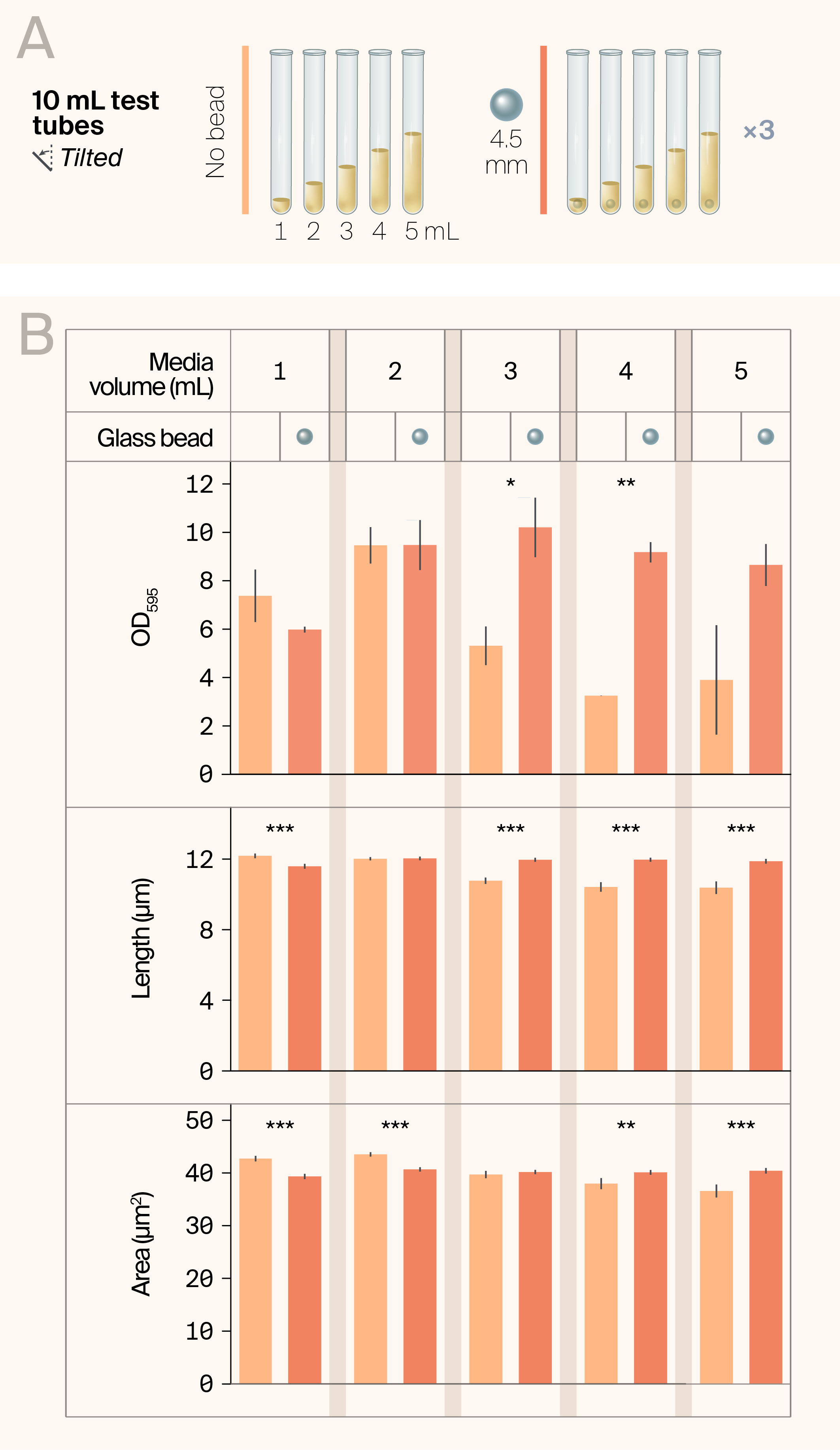
Figure 2. Beads promote the growth of large cultures in test tubes.
(A) We grew cultures of S. pombe overnight in test tubes in a rack placed in a tilted rack holder, with three biological replicates per strain and treatment.
(B) Average phenotypic measurements for each growth condition. Error bars represent 95% CI, with N = the # of cells imaged. Statistical differences between cultures with vs. without a bead are indicated with asterisks: *p < 0.05, **p < 0.01, ***p < 0.001 (Welch's t-tests).
Measuring cell growth at OD595, we found that culture volume had a large effect on how cultures responded to beads (Figure 2, B). At 1 mL, beadless cultures grew more densely than beaded cultures, although the difference wasn't statistically significant. At 2 mL, they grew similarly with beadless cultures showing initial signs of sedimentation. At 3–5 mL, beaded cultures grew to similar densities as at 2 mL. Still, beadless cultures showed widespread sedimentation and significant decreases in culture density (high variability in the 5 mL beadless cultures led to loss of statistical significance compared with the beaded 5 mL cultures). The most concentrated cultures in test tubes for both strains were at 3 mL at 10.2 OD595 (Figure 2, B). Because OD measurements are calculated using an equation that underestimates ODs above ~7 OD, it’s very likely that ODs reported here are slightly higher. These results suggest that yeast grown in volumes above 1 mL in a test tube will grow to a consistent and relatively high density with a bead.
While growing overnight cultures with beads improves culture density, we wanted to ensure that this increased density didn’t result from bead-induced cell lysis or come at the expense of log-growth phenotypes. Encouragingly, microscopy images (available on Zenodo) didn’t show cell fragments. Across all culture volumes, beaded cultures exhibit consistent length and area phenotypes, while beadless cultures exhibit decreased length and area as culture volume increases (Figure 2, B). At 1 mL, beadless cells were longer and larger, matching the generally denser cell culture phenotype, suggesting that beadless cultures are better suited for exponential growth at 1 mL (Figure 2, B). In 2 mL cultures, the area was larger in beadless cultures, but length phenotypes were indistinguishable across cultures. In 3 mL cultures, the beaded cultures show longer cells but similar areas. Only in 4–5 mL cultures are cells reliably longer and larger in beaded cultures than in beadless cultures (Figure 2, B). Overall, both area and volume phenotypes showed much higher consistency across volumes when beads were present, whereas beadless cultures showed increasingly stationary phenotypes as culture volume increased. These results suggest that beads should be used at culture volumes above 1 mL in test tubes to produce consistently dense, long, and large cells.
Finally, we wondered whether increases in cell density within test tubes with beads might be partially due to a higher evaporation rate, resulting in smaller culture volumes post-incubation. We measured the volumes of cell cultures with and without beads to test for differences in evaporation. We found average volume losses of 0.12 mL and 0.13 mL in beaded and beadless cultures, respectively, suggesting no significant increases in evaporation due to bead presence.
The same cultures behave differently in 24-well plates
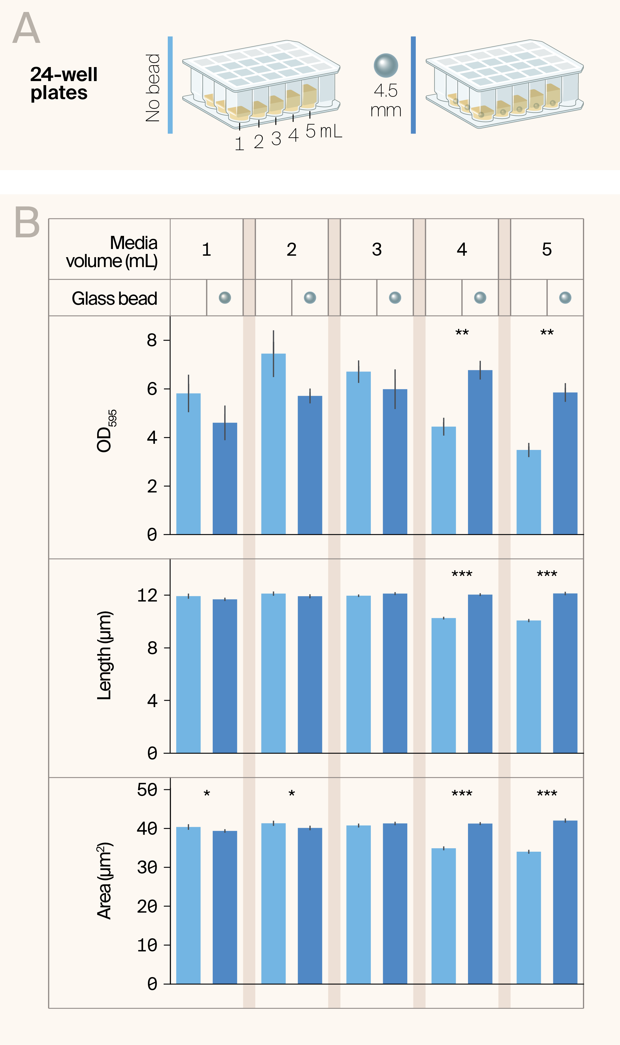
Figure 3. Beads have different effects on the growth of cultures in 24-well plates.
(A) We grew cultures of S. pombe overnight horizontally (not on a tilted base like in other experiments) in 24-well plates with three biological replicates per strain and treatment.
(B) Average phenotypic measurements for each growth condition. Error bars represent 95% CI, with N = the # of cells imaged. Statistical differences between cultures with vs. without a bead are indicated with asterisks: *p < 0.05, **p < 0.01, ***p < 0.001 (Welch's t-tests).
The shape of a container can significantly affect how liquids mix. For this reason, we ran the same tests in 24-well plates to compare with test tubes (Figure 3, A). The wells in 24-well plates are wider than test tube wells and square, promoting more turbulent mixing of cultures. At 2–3 mL, beadless wells showed a small amount of sedimentation, while beaded wells showed no sedimentation. At 4–5 mL, sedimentation was widespread in beadless wells. We generally found growth to be lower across all wells compared to test tubes, suggesting test tubes are preferable for growing high-density cultures across all tested perturbations (Figure 1, B). This time, while beaded cultures were again more consistent in density than beadless cultures, they were generally more inconsistent across volumes. The maximum OD595 of 7.5 was achieved in beadless 2 mL cultures. 4–5 mL beaded cultures were the only ones that were statistically significantly different, with beads consistently improving culture density at these volumes. The lower densities of 1–3 mL beaded cultures weren't statistically significant. These results suggest that beads help maintain culture density at high volumes but may be inconsequential or even detrimental at lower volumes.
When examining cellular phenotypes, area and length showed similar patterns. Both phenotypes were much more consistent in beaded wells than in beadless wells, with beadless wells showing a sharp drop-off in cell length and area at 4–5 mL volumes. At 1–3 mL volumes, cell phenotypes were indistinguishable with or without beads, with only marginally significant improvement in cell area at 1–2 mL beadless cultures (Figure 3, B). These high-volume cultures showed noticeable decreases in cell length and area compared to their beaded counterparts, likely due to more cells being in the stationary phase within the pellets. Thus, including beads in higher-volume 24-well plate wells is the best approach.
Taking all results into account, growing cells in a 24-well plate with beads produced consistent log-growth cell and culture phenotypes while preventing sedimentation. However, beaded cultures had lower, albeit statistically insignificant, densities at 1–3 mL volumes, suggesting beads aren't beneficial at lower volumes. Altogether, beads make phenotypes more consistent and increase culture density at larger volumes, much like they do in test tubes.
1 mL cultures grown in 96-well plates perform better with a bead
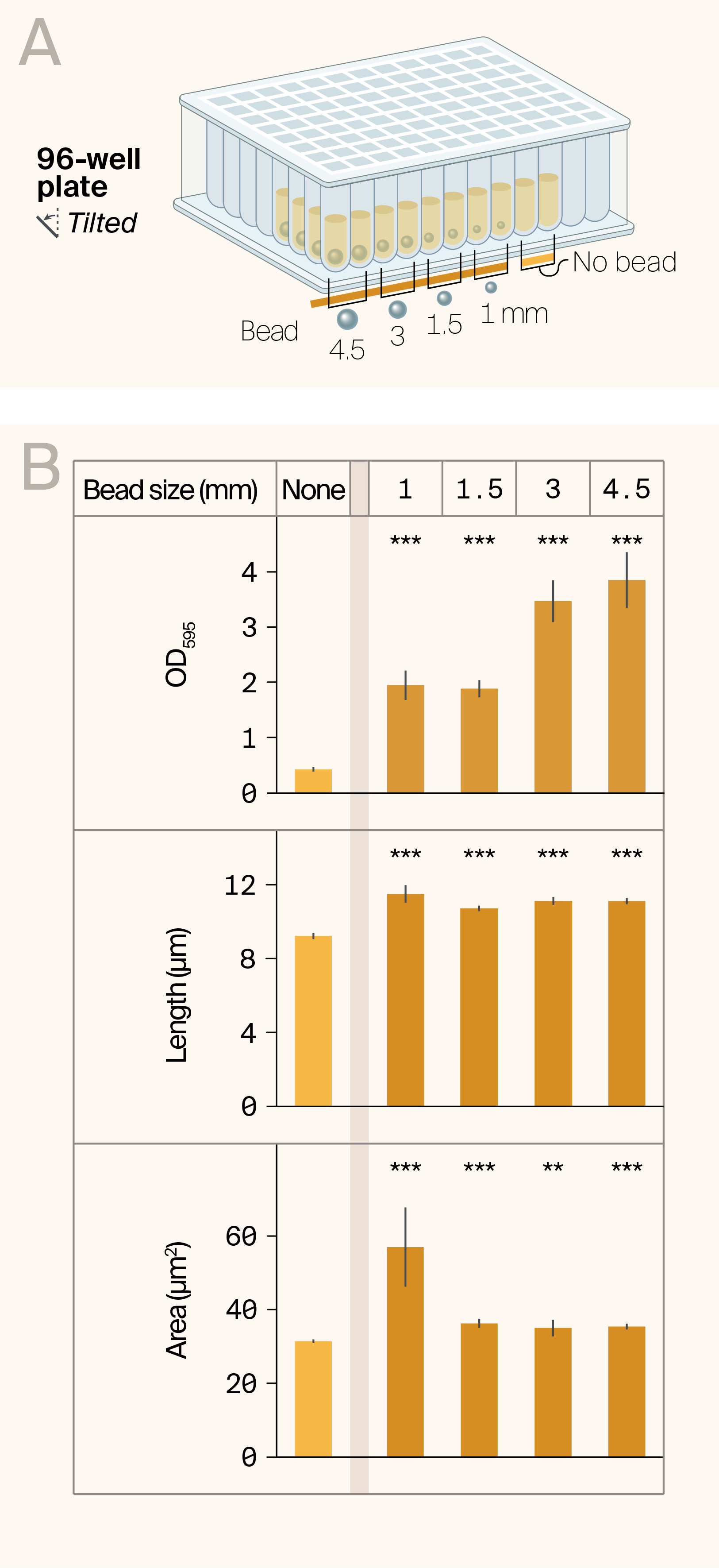
Figure 4. Large beads strongly promote culture growth in 96-well plates.
(A) We grew cultures of S. pombe overnight in 96-well plates and tilted on a plate holder, with eight biological replicates per strain and treatment.
(B) Average phenotypic measurements for each growth condition. Error bars represent 95% CI, with N = the # of cells imaged. Statistical differences between cultures with vs. without a bead are indicated with asterisks: *p < 0.05, **p < 0.01, ***p < 0.001 (Welch's t-tests).
While 24-well plates are useful for higher-throughput culturing, finding a reliable way to grow cultures in 96-well plates is critical for carrying out even higher-throughput culturing. That said, 96-well plates provide the biggest challenge. Their wells are very narrow and small in volume, both of which prevent uniform mixing of cultures and the growth of large volumes, where beads perform best in other containers. While 4.5 mm beads fit in these wells, they fit very snugly, potentially restricting their movement and, in turn, their ability to create turbulence in the wells.
To optimize the growth of yeast cultures in 96-well plates, we tested the effects of including different-sized beads or no beads at all (Figure 4, A). When we measured OD values of cultures after 2 days of overnight incubation at an angle, all beaded cultures showed clear, strongly significant increases in density compared with beadless cultures (Figure 4, B). Surprisingly, cultures showed increasing cell densities with increasing bead sizes, with 4.5 mm beads producing the densest cultures, around ten times denser than beadless cultures (Figure 4, B). These results mirror the fact that sedimentation was widespread in beadless cultures but decreased with increasing bead size. 3 mm and 4.5 mm bead-size cultures showed no visible sedimentation. This suggests that, unlike in wider vessels, 4.5 mm beads are beneficial in producing dense 1 mL cultures in 96-well plates.
When testing length and area phenotypes, differences between beaded and beadless cultures were measurable (Figure 4, B). Together, these experiments point to 96-well plate cell cultures benefiting the most from the addition of 4.5 mm beads.
Conclusion: Beads reliably promote logarithmic growth across vessels and volumes
After all these tests with different vessels and volumes, we wondered if there was a clear winner. Which growth vessel and media volume yield the densest, most homogeneous cultures? The answer depends on your experimental needs and capabilities. After testing more than 24 different experimental culture conditions, beaded test tube cultures are the clear winners, achieving the highest culture densities, the largest cells, and the longest cells. The data suggest that cultures with volumes greater than 5 mL may continue to show improvements, as indicated by an upward trend between culture volume and cell size and length (Figure 5, A–C).
However, 96-well plates are the best choice when working with large numbers of cultures. 96-well plates are the most space-efficient and the most compatible with laboratory robotics equipment and multi-channel pipettes. They allow more parallel experiments to be run simultaneously while still producing dense cell cultures with clear log-phase growth phenotypes. When compared directly to other cultures in other containers, beaded 96-well cultures grow to lower densities, suggesting they'll take longer to reach higher densities (Figure 5, A). Also, cells grown in 96-well plates are slightly shorter but have areas indistinguishable from those of cells grown in other containers (Figure 5, B & C). Together, these suggest that beads allow cultures in 96-well plates to come much closer to the quality of cultures in other containers, making cell culturing viable in 96-well plates.
24-well plates don’t show a clear benefit above the others in either direction. However, they might work as the best of both worlds. They produce consistent log-growth phenotypes, similar to test tubes. Still, they’re compatible with some high-throughput workflows without risking overgrowth or slight reductions in cell length and area that may occur in 96-well plates (Figure 5, A–C).
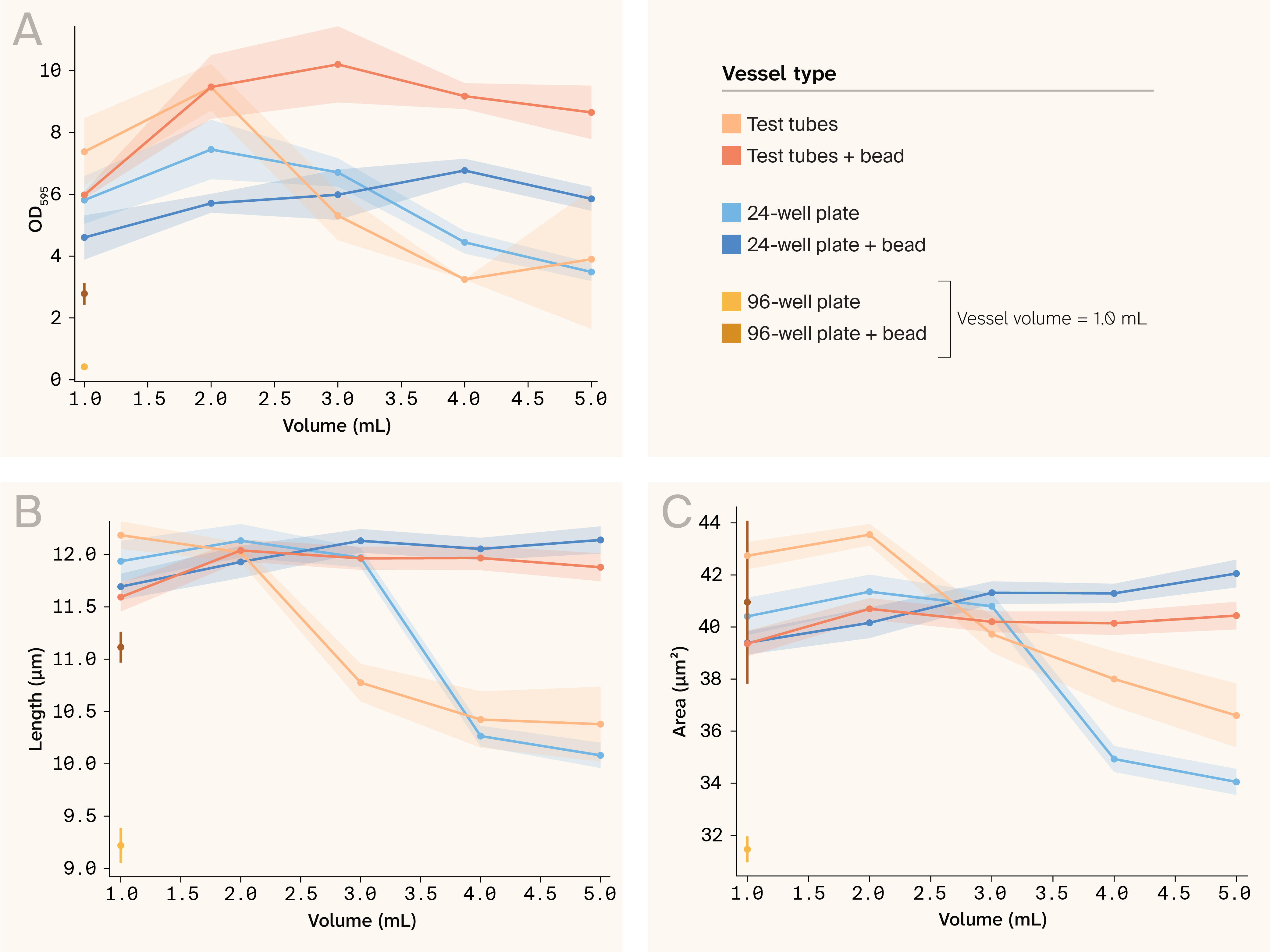
Figure 5. Cultures across vessels generally benefit from the addition of beads.
We’ve plotted average phenotypic data from cultures we grew in different vessels, with and without 4.5 mm beads. We facet the data by strain: wild-type on the left, mutant on the right. For each, we compare the average value of four phenotypes: (A) OD595, (B) length, (C) area measurements. Error bars represent 95% CI.
Bonus data: Dextrose and vitamin supplementation of media can improve culture densities without phenotypic effects
While not part of the main bead story, this section may be useful to those interested in growing S. pombe with supplemental nutrients.
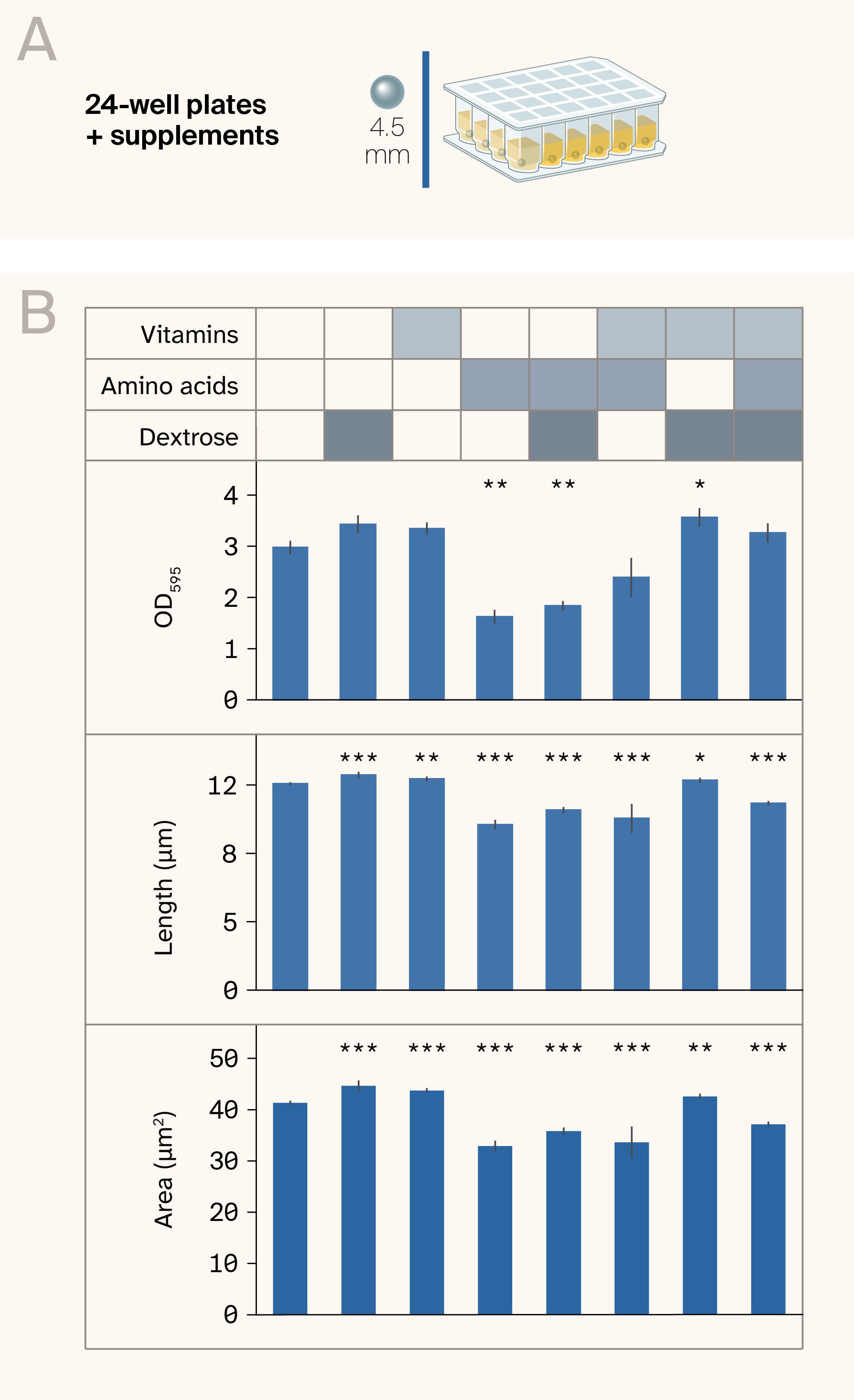
Figure 6. Supplementation with vitamins and dextrose creates denser cultures.
(A) We grew cultures of S. pombe overnight in 24-well plates with a bead in each culture, with three biological replicates per strain and treatment.
(B) Average phenotypic measurements for each growth condition. Error bars represent 95% CI, with N = the # of cells imaged. Significant differences between cultures with vs. without supplementation are indicated with asterisks: *p < 0.05, **p < 0.01, ***p < 0.001 (Welch's t-tests).
Would nutritional supplementation of minimal media further improve the growth rate and cellular phenotypes of S. pombe? We found a small yet statistically significant promotion of logarithmic growth phenotypes in cultures supplemented with dextrose and vitamins.
Stationary-phase phenotypic changes are a protective cellular response to nutrient deprivation that helps cells survive. Multiple studies have looked into the effects of nutrient availability on cell phenotypes 6–7. They suggest that amino acids, vitamins, and sugar can affect cellular phenotypes and help boost culture growth. We used the supplements mentioned in these studies, as well as common supplements added to different versions of EMM media, to see if they'd improve phenotypes associated with nutrient depletion.
We tested a few supplementation options in our beaded, 5 mL 24-well plate cultures clustered into three groups (Figure 6, A): vitamins, amino acids, and sugar (3% dextrose). Both vitamin and dextrose supplementation increased culture density, as measured by OD values (Figure 6, B). Their effects were somewhat additive, with density improvements being statistically significant only in dextrose-vitamin-supplemented media. When we checked cells under the microscope, we found dextrose and vitamins, both alone and together, statistically increase cell length and area (Figure 6, B). Surprisingly, the addition of amino acids resulted in cell cultures that were significantly less dense than unsupplemented cultures, and in cells that were shorter and smaller (Figure 6, B). That said, media supplemented with amino acids + vitamins + dextrose partially rescued the detrimental growth and phenotypic effects of amino acid supplementation (Figure 6, B).
Together, these experiments suggest that adding dextrose and vitamins can further increase the expression of exponential growth phenotypes, albeit only marginally.
Key takeaways
Adding a single bead to a range of culture volumes and containers promoted culture growth and made phenotypes much more consistent. Adding beads prevents cell sedimentation, a common problem in growing many different species, and one with significant effects on cell quality. Based on our findings, we'd recommend adding a single bead to most S. pombe cultures.
Specific findings:
- Beads prevented sedimentation and reduced phenotypic differences across culture volumes.
- In most cases, cultures grown with beads reached higher ODs faster, and showed increased cell size and length.
- To our knowledge, this is the first observation that beads can be beneficial — not stressful — to S. pombe cells in culture.
- Beads allow consistent growth of S. pombe cells across all the vessels we tested, with test tubes producing the densest cultures and 96-well plates showing the greatest improvement in cell length, cell area, and culture density phenotypes.
- Adding beads may sufficiently support cell growth, allowing experimenters to replace multiple baffled flasks with 96-well or 24-well plates, providing space-saving and automation benefits that enable high-throughput experimental workflows.
Importantly, our experiments show that the vessel and volume in which cell cultures grow significantly impact their phenotypes and densities, underscoring the need to document these when controlling for growth conditions. Most published methods fail to provide details about the containers used to grow their cell cultures. Our findings suggest that reporting the culturing containers can improve the reproducibility of your results.
Next steps
Test beads in growing different species
As of the time of this publication, other scientists at Arcadia have tested the effects of beads on the growth of Chlorella vulgaris, Saccharomyces cerevisiae, and Chlamydomonas reinhardtii in liquid cultures. So far, they’ve found that all have benefited from growth with beads in at least one of the tested conditions — stay tuned for detailed data in future pubs. However, under certain conditions, adding beads can result in visible cell fragments in algal cell cultures, suggesting that beads can indeed lyse cells at higher growth temperatures or at higher rotational speeds during incubation.
What cellular characteristics could help us predict which species and strains of cells are most likely to benefit from the addition of beads to culture? For species that'll likely show improved culture quality with beads, in what conditions are those beads best able to promote culture quality? We’re looking into these questions, and we encourage others to do the same.
Try new bead materials
In some vessels and at low culture volumes, adding beads can be stressful to cells. Glass and zirconium beads can create friction and are used to help lyse cells. We're interested in testing other materials, such as silicone and agarose beads, to see whether lighter, more flexible beads can provide better growth conditions that reduce cell stress. These beads might be especially important for the growth of S. pombe strains with weak cell walls or for species without cell walls. We're also testing stainless steel beads that are magnetic to see if using magnets can make bead retrieval after culture growth easier, while promoting culture growth without inducing significant cell stress.
Vary the shaking speed and angle
Commercially available test tube racks for growing cell cultures in shaking incubators are commonly designed to accommodate growth at different angles. This suggests that growing tilted cultures has a positive effect on culture growth and is a major reason for the tilted holders we used in this publication. However, most manufacturers don't specify which tilt angles to use for culturing or how tilt angle affects cell growth, and we didn't test different tilt angles in our experiments. Preliminary experiments in C. vulgaris and C. reinhardtii suggest that, at least in 96-well plates, the tilt angle has significant effects on growth. Documenting how the tilt angle of cultures in test tubes and 96-well plates affects growth would help inform decisions on culturing angles for different cells.
While there’s extensive information available on the effects of rotational speed on the growth of cultures 8–9, it's likely to have different effects on beaded cultures, at different angles, and in different vessels. We plan to test different angles and rotational speeds for cell cultures and report our findings to help other scientists boost their cell cultures.
Future applications
Moving forward, we're likely to test beads to grow other organisms at Arcadia, especially when we use 96-well plates, and will continue to share what we find.
Weigh in!
We're eager to hear from other scientists. Does adding beads to your organism’s cultures help mitigate issues with cell sedimentation, improve growth rates, or produce more consistent cellular phenotypes? Please comment here if you give beads a try!
Acknowledgements
Acknowledgements
We'd like to thank Hayley Greenough for providing liquid cultures and plates used in this study.
Be the first to comment on this publication.